Chest Pain X-ray
Date: 2026-07-28
Accepted answers: Coarctation of aorta
Explanation
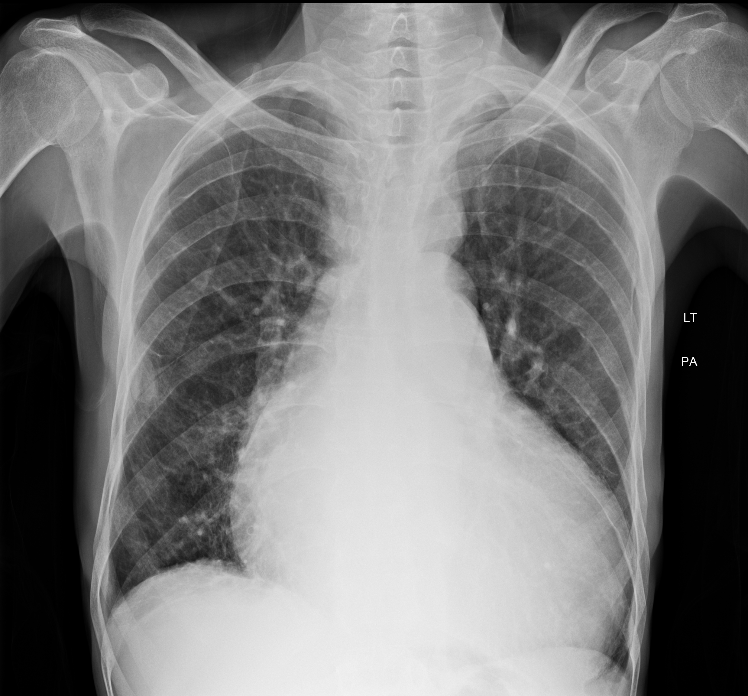
Two findings carry the diagnosis on this film. The indentation at the aortic isthmus with pre- and post-stenotic dilatation produces the figure-of-3 sign, and dilated intercostal collaterals erode the inferior rib margins, sparing ribs 1 and 2 because the costocervical trunk supplies them rather than the internal mammary collateral pathway. Chronic afterload drove the LV hypertrophy and eventual failure seen here.
Source: Patil S, Coarctation of aorta. Case study, Radiopaedia.org (Accessed on 28 Jul 2026) https://doi.org/10.53347/rID-223566
Hints
- A 35-year-old man presents with several months of progressively worsening exertional dyspnea, dizziness, fatigue, and chest pain.
- Blood pressure measures 178/96 mmHg in both arms and 118/70 mmHg in the lower extremities. In addition, femoral pulses are weak and delayed relative to the radial pulses, and a systolic murmur radiates to the left interscapular area.
- ECG shows left ventricular hypertrophy with a lateral strain pattern, and he reports years of documented hypertension without prior cardiac workup.
- Frontal chest radiograph shows massive cardiomegaly with a cardiothoracic ratio of 0.75 and cephalisation of the left upper lobe pulmonary veins. There is no pleural effusion, hilar enlargement, or pruning of the pulmonary vessels.
- The lesion occurs in two forms being preductal (infantile, typically a long hypoplastic segment) and postductal (adult, typically focal).
- The aortic contour is abnormal, with a focal indentation at the isthmus flanked by dilatation above and below. Bilateral inferior rib notching involves the posterior third through eighth ribs and spares the first two.