Something looks to big X-ray
Date: 2026-04-25
Accepted answers: Pericardial effusion
Explanation
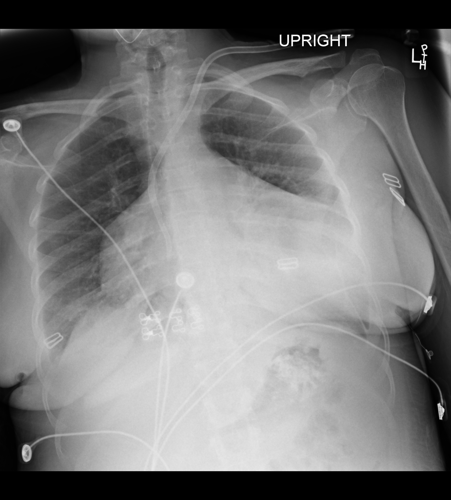
The chest radiograph shows a markedly enlarged, globular cardiac silhouette with a classic water-bottle configuration, reflecting a large pericardial effusion; notably, pulmonary vascularity is preserved, a key feature distinguishing pericardial effusion from congestive heart failure, where pulmonary venous congestion and interstitial edema would be expected. In uremic patients, inflammation of the pericardial lining leads to fluid accumulation within the pericardial space, and when this occurs rapidly, even moderate volumes can raise intrapericardial pressure enough to impair ventricular filling. A condition known as cardiac tamponade, clinically identified by Beck's triad of hypotension, elevated JVP, and muffled heart sounds. Echocardiography is the confirmatory study of choice and is essential for assessing the size and hemodynamic significance of the effusion before drainage. Management includes urgent pericardiocentesis when tamponade physiology is present, while intensification of hemodialysis can resolve uremic effusions in hemodynamically stable patients.
Source: Aliyari M, Pericardial Effusion. Case study, Radiopaedia.org (Accessed on 25 Apr 2026) https://doi.org/10.53347/rID-234347
Hints
- A 40-year-old woman with end-stage renal disease on maintenance hemodialysis presents to the emergency department with progressive dyspnea over the past several days.
- On physical examination, she is mildly hypotensive, with markedly elevated jugular venous pressure and muffled heart sounds on auscultation.
- She reports missing several recent dialysis sessions, and laboratory workup confirms severe uremia with a BUN greater than 100 mg/dL; ECG shows diffuse low-voltage complexes across all leads.
- Chest radiograph demonstrates marked global enlargement of the cardiac silhouette with a smooth, rounded "water bottle" contour, without redistribution of pulmonary vasculature or interstitial edema to suggest volume overload.
- This complication occurs in up to 20% of patients on chronic hemodialysis and is driven by uremic inflammation of the pericardium; when it develops rapidly, even modest fluid accumulation can precipitate life-threatening hemodynamic compromise.
- A hemodialysis catheter tip is visible projecting over the right atrium; the cardiac silhouette enlargement is smooth, symmetric, and globular with preserved lung fields, reflecting fluid accumulation within the fibrous sac surrounding the heart, a finding best confirmed and mapped with bedside ultrasound prior to drainage.