Pain in the Knees X-ray
Date: 2026-03-11
Accepted answers: Osteochondroma
Explanation
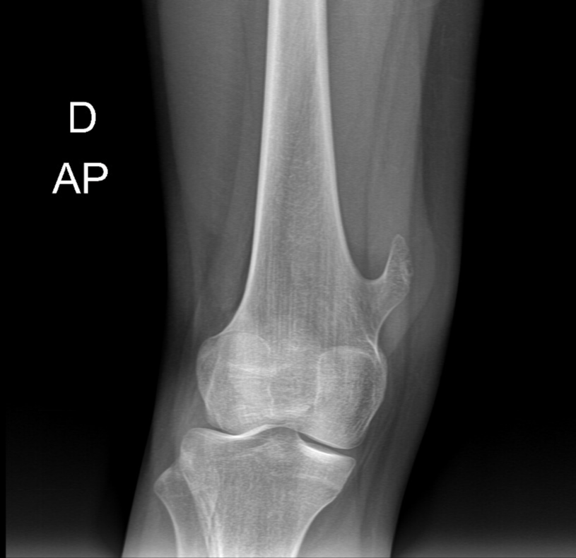
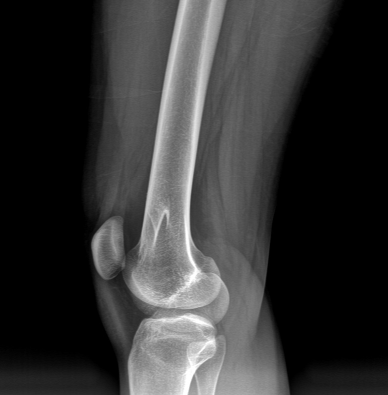
Osteochondromas are the most common benign bone tumors, typically arising during the second decade of life as the skeleton is still growing. They originate from the metaphyseal growth plate cartilage and consist of a cortical and medullary component that is continuous with the host bone. The pedunculated variant (as seen in this case) projects away from the metaphysis, often near the distal femur, proximal humerus, and proximal tibia—areas of rapid skeletal growth. While most osteochondromas remain asymptomatic and require no treatment, symptomatic lesions causing mechanical irritation of adjacent soft tissues may warrant surgical excision. Malignant transformation, though rare, is a concerning long-term complication; however, the absence of a thickened cartilaginous cap and stable imaging appearances after skeletal maturity effectively excludes this risk. Management is conservative with clinical and radiographic surveillance.
Source: Lustosa L, Osteochondroma. Case study, Radiopaedia.org (Accessed on 11 Mar 2026) https://doi.org/10.53347/rID-230933
Hints
- A 15-year-old female presents with a hard, palpable mass over the right knee that has been present for several months and is growing.
- The mass is fixed to bone, painless on direct palpation, and causes mild discomfort only with certain knee movements and activities. There are no constitutional symptoms, fever, or systemic signs of malignancy.
- On plain radiographs, a bony projection is identified arising from the metaphyseal region of the distal femur. The lesion is pedunculated with a stalk-like appearance and a broad base of attachment to the underlying bone.
- The lesion demonstrates continuous cortical and medullary bone with the host skeleton, with no periosteal reaction, cortical disruption, or underlying bone destruction. The growth is directed away from the joint space.
- Radiographic features confirm mature osseous composition with a well-defined, sharply marginated transition zone and no evidence of soft tissue ossification, aggressive features, or malignant changes such as peripheral calcification or a thick radiolucent cap.
- This benign metaphyseal lesion arises from growth plate cartilage and typically remains stable after skeletal maturity. The absence of mechanical symptoms or imaging signs of malignant transformation argues against surgical intervention at this time; surveillance imaging is appropriate.