Pain in the Hips X-ray
Date: 2026-03-05
Accepted answers: Osteoarthritis
Explanation
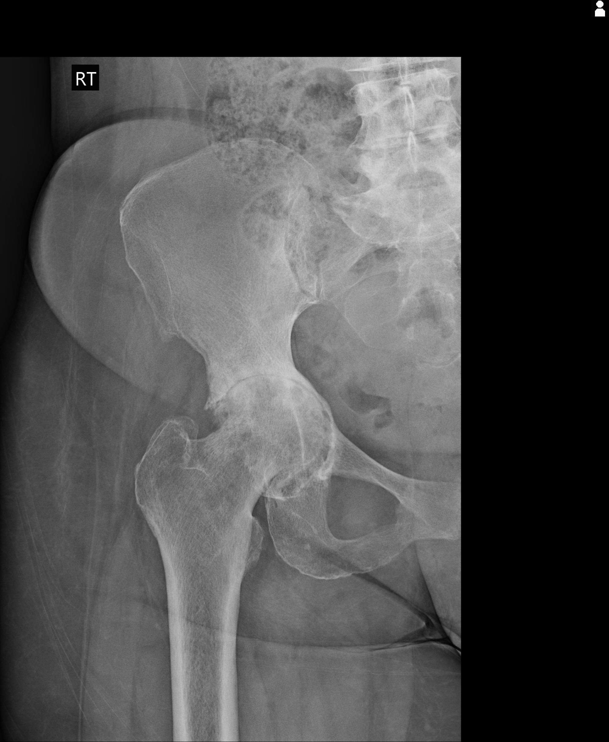
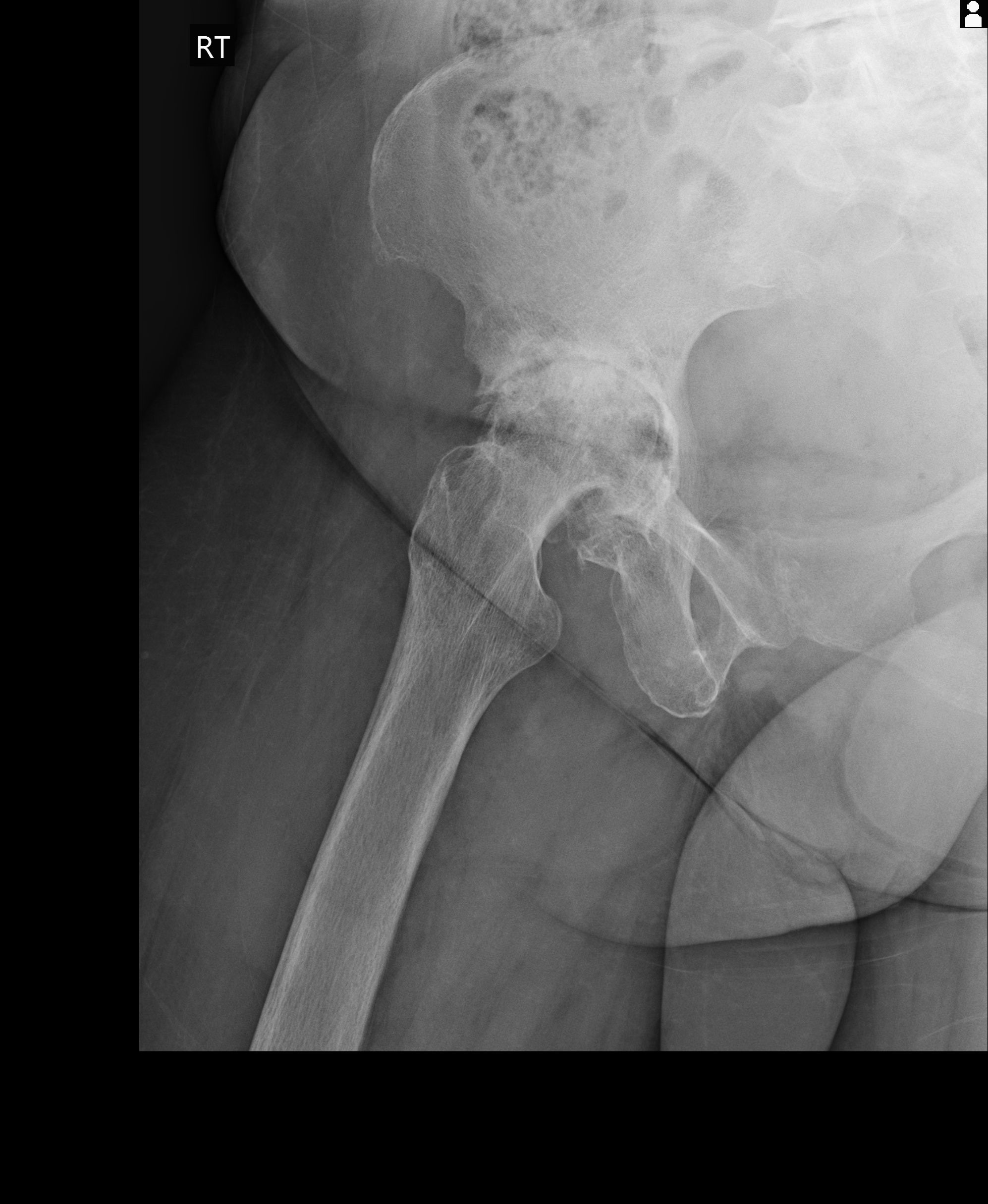
These radiographs demonstrate advanced degenerative joint disease of the right hip (Kellgren-Lawrence grade 4). Key findings include severe joint space narrowing, subchondral sclerosis and cyst formation, marginal osteophytes, and acetabular protrusio where the femoral head migrates medially through the acetabular wall. This constellation of findings in a 70-year-old woman with mechanical pain patterns is classic for primary osteoarthritis, though secondary causes and inflammatory arthropathies should be considered in the differential given the severity and presence of protrusio.
Source: Lawal A, Advanced right hip osteoarthritis. Case study, Radiopaedia.org (Accessed on 05 Mar 2026) https://doi.org/10.53347/rID-229831
Hints
- A 70-year-old woman presents to the clinic with chronic right hip pain that has gradually worsened over several years.
- The pain is worse with weight-bearing activities and at the end of the day, with some relief at rest. She reports morning stiffness lasting less than 30 minutes.
- Physical examination reveals reduced range of motion in the right hip, particularly with internal rotation and abduction, along with crepitus on movement.
- Radiographs demonstrate marked narrowing of the right hip joint space with loss of the normal cartilage interval between the femoral head and acetabulum.
- The imaging shows subchondral sclerosis and multiple cystic changes in both the acetabulum and femoral head, along with marginal osteophyte formation at the acetabular rim.
- The femoral head is displaced medially beyond the ilioischial line, indicating protrusion into the pelvis, a finding more commonly seen in chronic inflammatory or degenerative processes.