The hands are in Pain X-ray
Date: 2026-03-02
Accepted answers: Giant Cell Tumor
Explanation
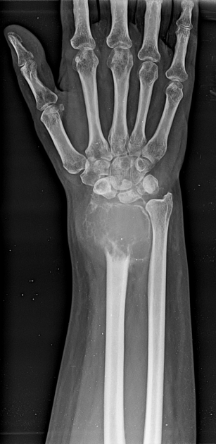
Giant cell tumor (GCT) is a benign but locally aggressive bone lesion that characteristically presents in young to middle-aged adults (20-40 years) with insidious, progressive pain and swelling at the distal femur, proximal tibia, or distal radius. The diagnosis is secured by the classic radiographic triad of epiphyseal-metaphyseal location with extension to the articular surface, purely lytic appearance with narrow zone of transition, and cortical expansion without periosteal reaction—as demonstrated in this case. GCT is histologically composed of multinucleated giant cells within a mononuclear stromal background and must be differentiated from aneurysmal bone cyst (younger patients, faster growth), brown tumor of hyperparathyroidism (biochemical abnormalities), and tuberculous osteomyelitis (systemic signs and endemic areas). MRI is the next appropriate imaging study to further characterize the lesion and guide surgical planning, which typically involves curettage or wide resection, though high recurrence rates (10-65%) are common with curettage alone. The absence of trauma history, fever, or constitutional symptoms combined with the imaging findings makes GCT the most likely diagnosis in this clinical scenario.
Source: Maitra D, Giant cell tumor of distal radius. Case study, Radiopaedia.org (Accessed on 02 Mar 2026) https://doi.org/10.53347/rID-213881
Hints
- 40-year-old female presents with gradually progressive pain and swelling of the right wrist over 3 months. No history of trauma or antecedent injury.
- No fever, no chills, no night sweats, and no unintentional weight loss. Constitutional symptoms are notably absent.
- Lesion is typical for arising in the epiphysis after physeal closure in young to middle-aged adults.
- Radiographs demonstrate a lesion at the distal radius, involving both the metaphysis and epiphysis with extension to the articular surface of the radiocarpal joint.
- The lesion is purely lytic with a narrow zone of transition. There is cortical expansion with thinning and focal breaches, but the overall bone architecture remains intact. No periosteal reaction, no soft tissue mass, and no sclerotic rim.
- The epiphyseal-metaphyseal location with extension to the articular surface narrows the differential significantly. This finding in a skeletally mature patient is relatively uncommon and helps exclude many other lytic bone lesions.