The Arm X-ray
Date: 2026-02-13
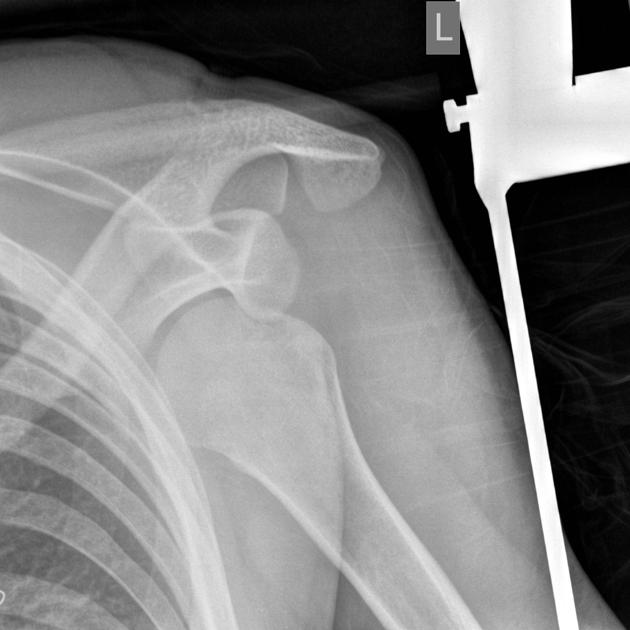
Accepted answers: Anterior Shoulder Dislocation
Explanation
When evaluating a shoulder radiograph for a suspected anterior dislocation, begin by confirming the view and ensuring image quality, making sure the clavicle, glenoid, and proximal humerus are adequately visualized. Next, assess alignment by determining whether the humeral head is centered over the glenoid fossa. In a normal shoulder, the humeral head should sit congruently within the glenoid; if it appears displaced medial and inferior to the glenoid, this suggests an anterior dislocation, most commonly a subcoracoid type. The glenoid may appear “empty,” reflecting loss of normal joint articulation. After identifying displacement, carefully inspect for associated injuries such as a greater tuberosity fracture, a Hill-Sachs lesion, or a Bankart lesion, as these commonly accompany dislocations. Correlating radiographic findings with the mechanism of injury, typically forced abduction and external rotation, strengthens diagnostic confidence. A systematic approach focusing on alignment first helps ensure accurate interpretation and reduces missed pathology.
Source: Jones J, Anterior shoulder dislocation. Case study, Radiopaedia.org (Accessed on 14 Feb 2026) https://doi.org/10.53347/rID-7132
Hints
- A 24-year-old male presents after a fall onto an outstretched arm while playing basketball.
- He reports severe shoulder pain and inability to move the arm. He is holding the affected arm slightly abducted and externally rotated.
- On exam, there is loss of the normal rounded contour of the shoulder with a visible prominence inferior to the clavicle.
- Injury occured with the arm forced into abduction and external rotation
- On AP radiograph the humeral head is displaced medial and inferior relative to the glenoid fossa
- The humeral head lies anterior to the glenoid, often positioned subarachnoid